Few weeks ago we covered macro photography and watching closely photos of small things is always amusing. This year Nikon again put up a contest and announced winners of Microphotography, where we will see things that we never have seen before. The vibrant colors and the close up of these lively things is really fascinating.
These photos might look like something out of the world but after reading their details you will be amazed as I am. NATGEO placed a page for the this photography as well do check it out. Microphotography have many techniques and we tried to write them down along with the image details.
Nikon Small World winners list and their images are below check them out.
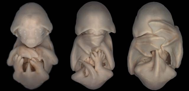
20 – Black Mastiff Bat
Embryos of the species Molossus rufus, the black mastiff bat. These images formed part of an embryonic staging system for this species. The technique to take images is called Brightfield.
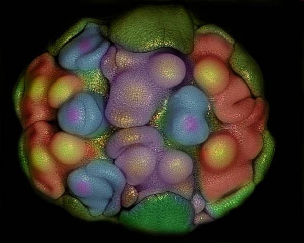
19 – Garlic
This is a Floral primordia of Allium Sativum (Garlic). Now one can know why vampires are scared of Garlic. The technique for this image is called “Epi-Illumination”.
18 – Coral sand
Coral sand is 100 times magnified and the imaging technique is called “Brightfield”.
17 – Leaf Vein
Stinging nettle trichome on leaf vein is magnified 100times and the technique for image is called Transmitted Light.
16 – Fossilized Turitella
Fossilized Turitella agate containing Elimia tenera (freshwater snails) and ostracods (seed shrimp) is magnified 7 times. The technque for this image is called “Stereomicroscopy”.
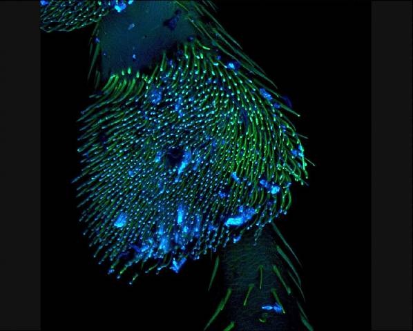
15 – Ladybug Leg
by Andrea Genre
Section of a Coccinella (ladybug) leg magnified for 10 times. The technique is called “Confocal”
14 – Pistil of Adenium obesum
Pistil of Adenium obesum magnified for 10 times. Technique is called “Image Stacking”
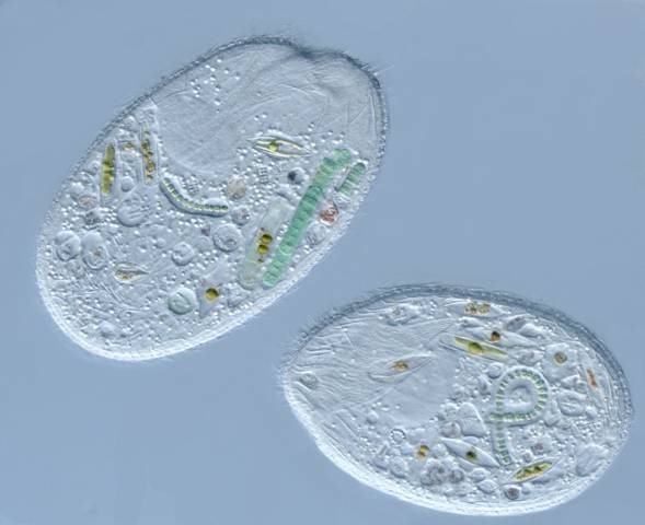
13 – Sonderia
Sonderia sp. (a ciliate that preys upon various algae, diatoms, and cyanobacteria) magnified for four hundred times. The technique is called “Nomarski Interference Contrast”
12 – Dextran Beads
by Esra Guc
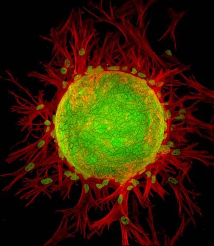
3D lymphangiogenesis assay. Cells sprout from dextran beads embedded in fibrin gel. Magnified for two hundred times. Technique for imaging is called “Fluorescence and Confocal”.
11 – Drosophila melanogaster larva
Single optical section through the tip of the gut of a Drosophila melanogaster larva expressing a reporter for Notch signaling pathway activity (green), and stained with cytoskeletal (red) and nuclear (blue) markers. Magnified for twenty five times and technique to take image is called “Confocal”.
10 – Brittle Star
A prickly looking brittle star was magnified eight times—and claimed tenth place—in this shot by Alvaro Migotto of Brazil’s University of São Paulo. The marine animal was photographed using a dark-field method, an illumination technique that creates a stark contrast between the specimen and the surrounding area. Technique for imaging is called Stereomicroscopy, Darkfield.
09 – Larva-Carrying Ant
by Geir Drange
Caught in the act of carrying its larva, an ant in the genus Myrmica—found throughout the Northern Hemisphere—is enlarged five times in this ninth-place. The technique for imaging is called Reflected Light, Image Stacking. Ants take their larvae in and out of the nest each day in order to regulate their temperature.
8 – Sea Gooseberry
This shot of sea gooseberry larvae was taken using technique called ‘Differential Interference Contrast’. Sea gooseberry larvae (Pleurobrachia) a type of ctenophore that’s 95 percent water—was magnified 500 times. It came in eighth place.
7 – Fruit Fly Eye
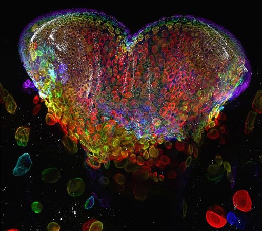
Magnified 60 times, the eye organ of a larval fruit fly (Drosophila melanogaster) is awash in surreal color. This image-which netted seventh place was made with a confocal lens. That makes different levels of the organ simultaneously visible. The technique of imaging is called Confocal.
6 – Vibrant Algae
by Marek Mis
This is a algal Cosmarium which was magnified one hundred times. The image won 6th place. The technique of imaging is called Polarized Light.
5 – Phosphate Closeup
Cacoxenite is a phosphate mineral that can range in color from yellow to brown to red. The University of Valencia’s Honorio Cócera-La Parra magnified this sample—collected from the La Paloma Mine in Valencia, Spain—18 times to capture the fifth-place prize. This image technique is “Transmitted Light”.
4 – Pupal Fruit Fly
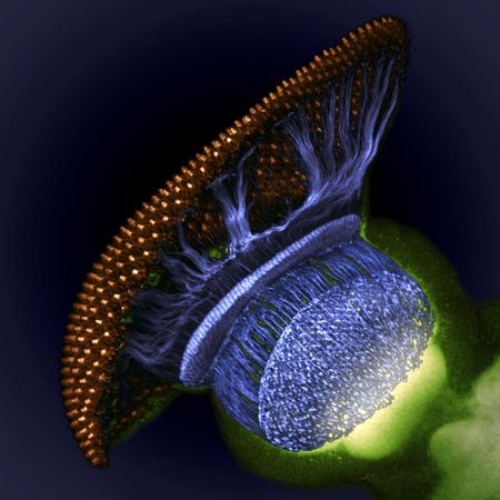
It’s not a spaceship. It’s the visual system of a pupal fruit fly (Drosophila melanogaster). Taken by W. Ryan Williamson of the Howard Hughes Medical Institute in Ashburn, Virginia, this shot claimed 4th place. This photo is 1500 times magnified. This image also uses “Confocal” technique.
3 – Bone Cancer Cell
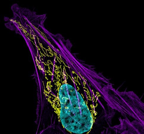
Human bone cancer showing actin filaments (purple), mitochondria (yellow), and DNA (blue). This image was 63 times magnified. The technique to take the image is called Structured illumination Microscopy (SIM).
2 – Baby Spiders
Photo of newborn Lynx spiderlings was captured by Walter Piorkowski and it was magnified 6 times. This photo earned 2nd place in the Nikon Small World contest. The image technique is “Reflected Light, Fiber Optics, Image Stacking”
1 – Bright Brain
by Dr. Jennifer L. Peters & Dr. Michael R. Taylor
This winning photo might look like sparkling lights but it’s a brain of Zebrafish embryo. This photo was taken by Dr. Jennifer L. Peters and Dr. Michael R. Taylor of St. Jude Children’s Research Hospital Memphis, Tennessee, USA. This shot was magnified 20 times. These images where first pseudo colored with a rainbow palette based on depth so that the color scheme would be informative and visually appealing. This first prize winner photo is considered to be the first ever photo to show the formation of the blood brain barrier in a live animal. This image taking technique is called “Confocal”











These really are out of this world images. Microphotography rocks…….